Functional Near-Infrared Spectroscopy (fNIRS) is a non-invasive brain imaging technique that measures changes in blood oxygenation and blood flow in the brain. It works by shining near infrared (NIR) light (700 900 nm) through the scalp and detecting how much light is absorbed by oxygenated (HbO) and deoxygenated hemoglobin (HbR) in the blood.¹

1 . Molina-Rodríguez, Sergio, et al. “Stress Estimation by the Prefrontal Cortex Asymmetry: Study on FNIRS Signals.” Journal of Affective Disorders, vol. 325, Mar. 2023, pp. 151–157, https://doi.org/10.1016/j.jad.2023.01.018. Accessed 11 Apr. 2023.
fNIRS measures brain activity by tracking changes in blood flow and oxygen levels. This is based on a process called neurovascular coupling, which is the way the brain responds to increased activity.
When neurons become more active, they need more energy, which comes from glucose and oxygen. To supply this, the body increases blood flow to the active brain region, delivering more oxygen through oxygenated hemoglobin (HbO). fNIRS detects these changes by shining harmless near infrared light on the scalp and measuring how much light is absorbed by the blood. Since oxygenated and deoxygenated hemoglobin absorb light differently, fNIRS can determine how oxygen levels are changing, helping scientists and doctors understand brain function.¹

1 . Molina-Rodríguez, Sergio, et al. “Stress Estimation by the Prefrontal Cortex Asymmetry: Study on FNIRS Signals.” Journal of Affective Disorders, vol. 325, Mar. 2023, pp. 151–157, https://doi.org/10.1016/j.jad.2023.01.018. Accessed 11 Apr. 2023.
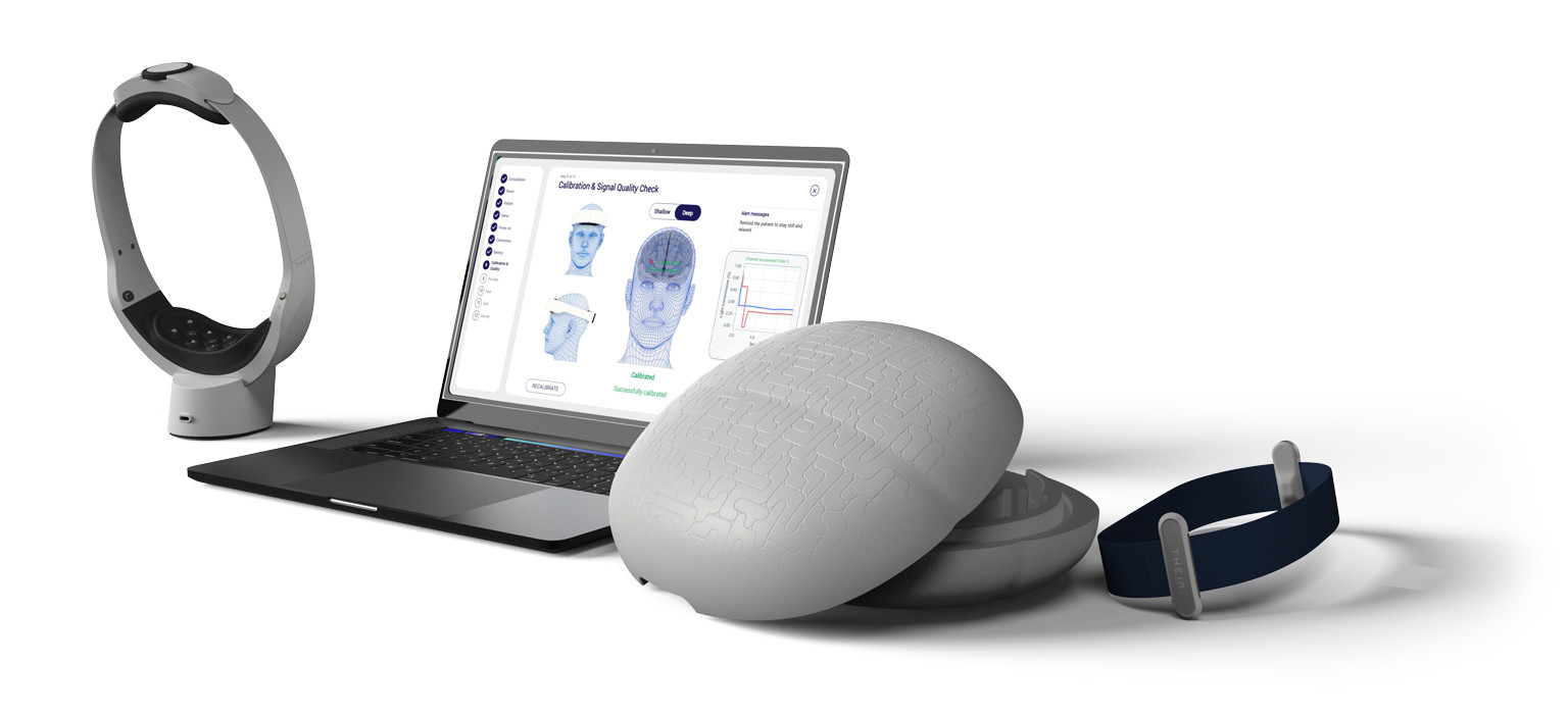
Theia analyzes brain hemodynamics by utilizing near-infrared (NIR) light, which can penetrate the scalp and reach blood vessels in the brain. Since biological tissues contain various chromophores (molecules that absorb light at specific wavelengths), it is crucial to differentiate those relevant to brain activity.
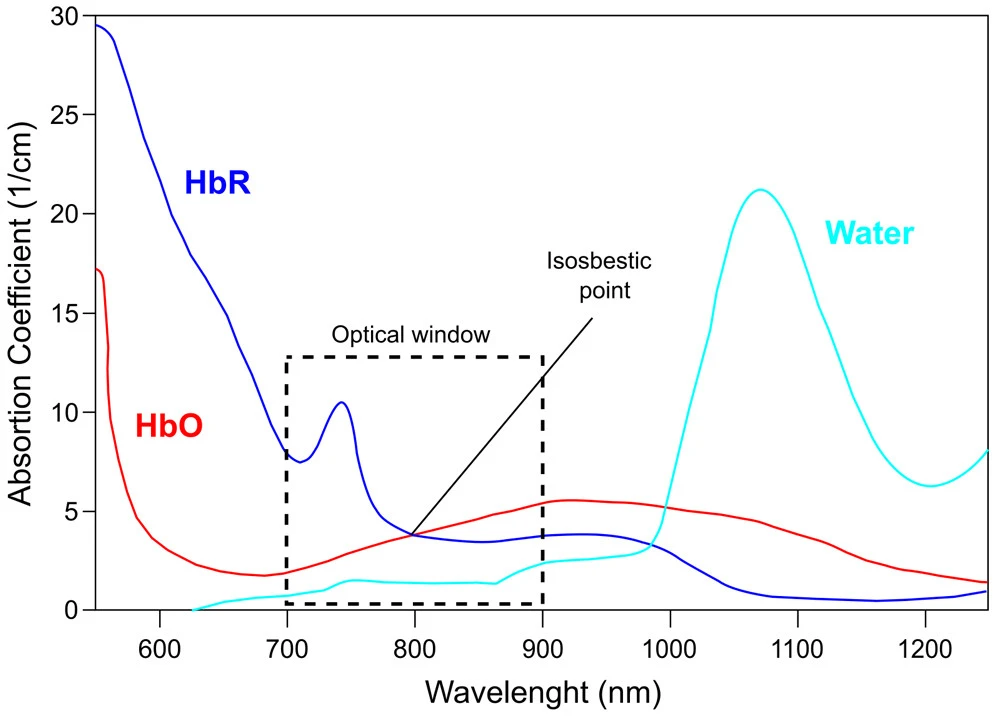
Water and lipids absorb very little light in the near-infrared (NIR) range (700–900 nm), rendering them largely invisible to the system. Theia focuses on hemoglobin, the keychromophore responsible for carrying oxygen in the blood, since it is the main absorber within the NIR optical window.¹
By emitting NIR light at optimized wavelengths (740 nm and 860 nm), Theia can distinguish between deoxygenated hemoglobin (HbR) and oxygenated hemoglobin (HbO). Since HbR absorbs more light at 740 nm and HbO absorbs more light at 860 nm, Theia detects the changes in light absorption at these specific wavelengths. By analyzing these variations, it can determine the relative concentration of HbO and HbR, allowing for real-time assessment of brain hemodynamics and oxygenation levels.
1 . Setchfield, Kerry, et al. “Relevance and Utility of the In-Vivo and Ex-Vivo Optical Properties of the Skin Reported in the Literature: A Review [Invited].” Biomedical Optics Express, vol. 14, no. 7, 21 June 2023, pp. 3555–3555, https://doi.org/10.1364/boe.493588. Accessed 29 Mar. 2024.
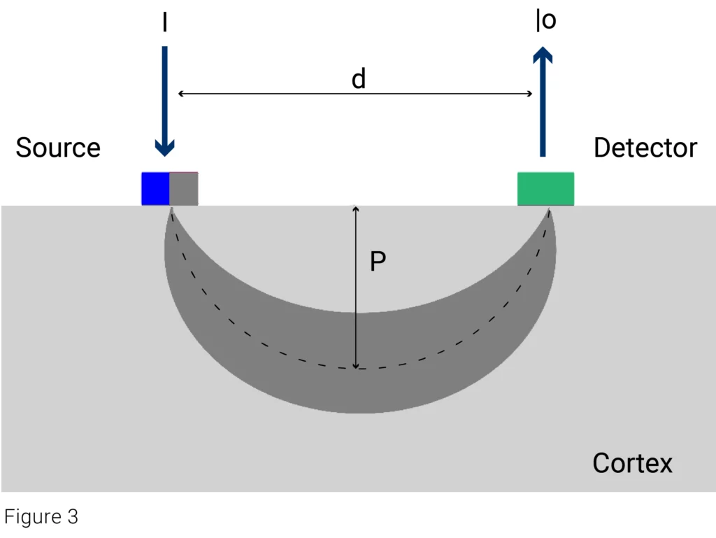
The propagation of light in tissues is governed by the absorption or dispersion of light photons. The photons follow an optical path that travels the “banana shaped” tissue from the light source (LEDs) to the detector (D). The depth that this banana-shaped path reaches in the cerebral cortex depends directly on the distance between LED and D.
The Lambert-Beer Law (also known as the Beer Lambert Law) describes how light is absorbed as it travels through a medium. It states that the amount of light absorbed depends on the concentration of the absorbing substance and the distance the light travels.
Mathematically, it is expressed as:
Oxygenation=CHBO -CHbR
- A = absorbance (how much light is absorbed)
- ε = molar extinction coefficient (how strongly the substance absorbs light at a specific wavelength)
- c = concentration of the absorbing molecule
- d = distance the light travels through the medium
fNIRS applies the modified Lambert-Beer Law to measure changes in oxygenated hemoglobin (HbO) and deoxygenated hemoglobin (HbR) concentrations in brain tissue over time.
fMRI provides very high spatial resolution and detailed images of the brain by detecting changes in blood oxygenation (the BOLD signal). However, it is expensive, less portable, and requires the subject to stay very still in a large, noisy scanner.
EEG records the brain’s electrical activity directly with very high temporal resolution (milliseconds), which is great for tracking rapid changes in neural activity. However, EEG has limited spatial resolution, making it hard to pinpoint exactly where in the brain the activity is occurring.
fNIRS offers a unique balance between cost, portability, and non invasiveness that sets it apart from fMRI and EEG.
Innovative techniques, such as salivary metabolite analysis and cerebrospinal fluid biomarkers, are being developed to differentiate between healthy individuals and those with dementia and to identify various types of dementia.
However, none of these tests are designed to distinguish between healthy individuals and those with MCI(an essential step for enabling early interventions to delay or prevent dementia) and they also rely on costly laboratory facilities and equipment.¹
1 .Botello-Marabotto M, Martínez-Bisbal MC, Calero M, et al. Non-invasive biomarkers for mild cognitive impairment and Alzheimer’s disease. Neurobiology of Disease. 2023;187(106312). doi:https://doi.org/10.1016/j.nbd.2023.106312
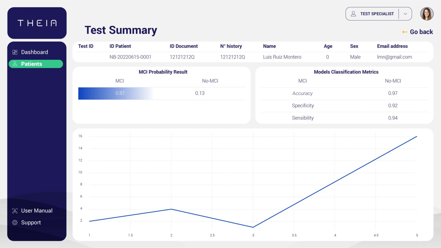
Studies carried out in Europe have shown a 92.6% AUC, 75% sensitivity and 94% specificity.¹